
Skin Spot Self-Checker: Suspicious Mole, Spot & Bump
Rixis Dermatology: Skin Spot Self-Checker
This tool is for educational purposes only and does not replace a professional medical evaluation. If you have any concerns about a skin lesion, please consult a board-certified dermatologist.
You will be asked a series of questions with example images to help compare your lesion’s appearance. We will suggest a possible concern and risk category – but only an in-person exam can confirm the diagnosis. We strongly recommend you see a dermatologist for proper evaluation or treatment regardless of our risk assessment.
Is your lesion mainly brown, black, or multi-colored?
A: Is your lesion asymmetrical?
B: Does your lesion have an irregular border?
C: Does your lesion show multiple or uneven colors?
D: Is your lesion larger than 6 mm?
E: Has your lesion changed recently (in size, shape, color, or started bleeding)?
Does your lesion have a waxy or “stuck-on” surface that can appear flakey or crumbly?
This can mimic nevi, including atypical nevi or melanoma.
Is the lesion scaly or crusted?
Does it feel thick, painful, or bleed with minor contact?
Sometimes this type of spot can appear raw or irritated, and bleed with slight pressure.
Does the lesion appear pink or red rather than matching your usual skin tone?
Look closely in good light to see if it has a pinkish, reddish, or sometimes reddish-purple tint, or if it is close to your normal skin color.
Does it have a shiny or pearly surface with small visible blood vessels?
Check for a glossy, translucent appearance with tiny branching vessels you can sometimes see with close inspection.
Is it a rapidly growing, firm bump that developed over weeks or months, sometimes red or purple?
Consider how quickly the lesion emerged. A sudden or fast growth that is firm and reddish to violet can be concerning.
Is it a bright red bump that may bleed easily if bumped?
Some spots are cherry-red or slightly darker and bleed readily with minor trauma.
Does it have a textured or bumpy surface or a central dimple?
Some lesions may look rough, like a small cauliflower (wart), or have a small dip in the center (molluscum).
Does it have a central dimple (umbilication) that you can see?
Molluscum contagiosum often has a distinct central depression. Warts usually lack that deep dimple.
Does it have a thin stalk (like a flap of skin) and move freely if you push it side to side?
Skin tags often appear in skin folds and can be easily moved around on a narrow base.
Is it a soft, mobile lump under the skin?
A lipoma often feels somewhat squishy and can move around when palpated.
Does it feel cystic (fluid-filled) with a visible pore or opening?
You might notice a somewhat softer center or a tiny opening at the top, unlike a firm, solid growth.
Do you have a personal/family history of skin cancer or intense sun exposure?
This tool does not replace professional care. We strongly recommend seeing a dermatologist for any lesion you are concerned about. A dermatologist can evaluate and treat skin conditions appropriately.
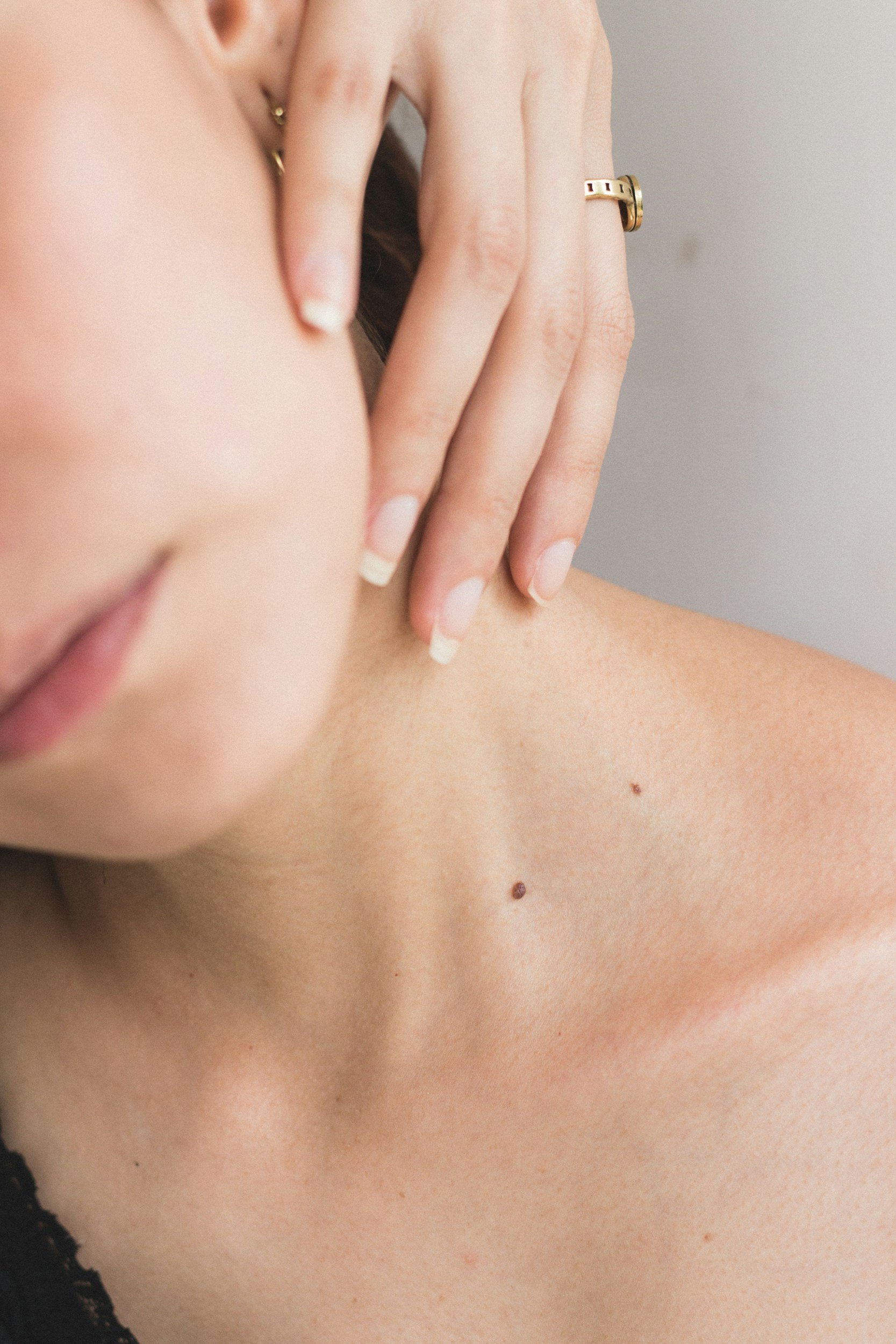
Concerned about a suspicious mole, spot, or unusual bump on your skin? At Rixis Dermatology in Columbus, Ohio, our free, educational Skin Spot Self-Checker may help you recognize common warning signs of skin cancer (including melanoma, basal cell carcinoma, or squamous cell carcinoma), as well as benign skin lesions such as warts, lipomas, or skin tags. This interactive tool uses a series of questions and example images to guide you in assessing the appearance of your lesion. Remember, this tool does not replace medical advice. Always follow up with a board-certified dermatologist or your healthcare provider if you have any concerns about your skin.
Why Use Our Skin Spot Self-Checker?
Identify Potentially Concerning Lesions: Learn to spot the ABCDE warning signs of melanoma and other types of skin cancer.
Educational & Easy to Use: This simple step-by-step approach can help you compare the appearance of your skin lesion with common examples.
Columbus, Ohio Dermatology Experts: Our team at Rixis Dermatology has extensive experience in diagnosing and treating a wide range of skin conditions.
When in Doubt, Seek Professional Evaluation: If your lesion looks suspicious, changes over time, or causes symptoms like pain or bleeding, schedule a dermatology appointment promptly.
Common Types of Skin Lesions to Recognize
Melanoma or Atypical Moles: Often appear as darkly pigmented lesions with asymmetry or irregular borders.
Basal Cell Carcinoma (BCC): May present as a pearly, shiny bump with visible blood vessels or a non-healing sore.
Squamous Cell Carcinoma (SCC): Frequently emerges as scaly, crusty patches or thickened lesions that can bleed easily.
Actinic Keratoses: Pre-cancerous lesions that are often rough, scaly, and sun-exposed.
Seborrheic Keratoses: Benign growths that often have a waxy, “stuck-on” look.
Warts: Rough, bumpy growths caused by the human papillomavirus (HPV).
Molluscum Contagiosum: Typically small, dome-shaped lesions with a central dimple (umbilication).
Lipomas: Soft, movable lumps of fatty tissue under the skin.
Skin Tags: Small, flesh-colored flaps of skin, often appearing in folds.
Cysts: Fluid- or keratin-filled bumps that can have a small pore or opening at the top.
10 Frequently Asked Questions
Below is an FAQ section that addresses queries commonly searched by individuals who suspect they have a suspicious skin lesion or want to learn more about using a self-check tool.
How do I know if a mole is suspicious?
A suspicious mole often follows the ABCDE criteria: Asymmetry, irregular Border, multiple or uneven Colors, Diameter larger than 6 mm, and Evolving (changing in size, shape, or color). Use our Self-Checker tool for guidance, but always see a dermatologist for a definitive diagnosis.Can the Skin Spot Self-Checker diagnose skin cancer?
No. Our Skin Spot Self-Checker is for educational purposes only. It can help you recognize warning signs, but an in-person biopsy or professional exam is the only way to confirm or rule out skin cancer.When should I see a dermatologist for a skin lesion?
If your lesion is growing rapidly, bleeding, itchy, painful, or meets any concerning criteria from the Self-Checker, schedule an appointment with a board-certified dermatologist. Early evaluation and treatment are vital for the best outcomes.Is a scaly patch on my skin always cancerous?
Not always. A scaly or crusty patch could be eczema, psoriasis, actinic keratosis, or squamous cell carcinoma. The Self-Checker can help narrow possibilities, but a professional dermatologic exam is key for an accurate assessment.What if my lesion isn’t brown or black—could it still be a skin cancer?
Absolutely. Basal cell carcinoma can appear pearly or pink, and squamous cell carcinoma can look scaly or crusted. Not all skin cancers are darkly pigmented. The Self-Checker includes pathways for both pigmented and non-pigmented lesions.Are there risk factors for skin cancer I should be aware of?
Yes. Family or personal history of skin cancer, significant sun exposure or sunburns, having numerous moles, and fair skin that burns easily are some examples. The Self-Checker will ask about risk factors to help highlight potential concerns.Does a "stuck-on," waxy growth mean I have cancer?
Not necessarily. A seborrheic keratosis can look like a waxy, stuck-on growth and is benign. However, it’s sometimes difficult to distinguish from melanoma. If uncertain, consult a dermatologist.How accurate is the tool if I’m not in Columbus, Ohio?
The tool is universally educational regardless of location. However, it does not replace a visit to a local board-certified dermatologist or a trusted healthcare provider near you—even if that’s not in Columbus, Ohio.What if I already have a dermatologist, but want a second opinion?
Getting a second opinion can be reassuring if you have concerns about a skin growth. Bring any previous biopsy results or clinical notes with you so the second dermatologist can make a well-informed assessment.Do I need a referral to see a dermatologist at Rixis Dermatology?
Some insurance plans require referrals, but most allow you to book an appointment directly. Please contact our office or check with your insurance provider to understand your specific plan requirements.